MainPage:Nuclear:Summer2016:PWO
PbWO4 Characterization and Recovery
Abstract
The potential of using Lead Tungstate crystals (PbWO4) in electromagnetic calorimeters like the Neutral Particle Spectrometer (NPS) at 12 GeV Jefferson Lab and in future projects such as particle identification in the endcaps of the EIC (Electron Ion Collider) detector has been researched extensively. The radiation hardness as well as the small Moliere radius of PbWO4 (PWO) crystals make them ideal for use in a compact detector. Additionally, the light yield of these crystals outperforms that of PbF2 and other heavy crystals, lending to even more sensitivity to event resolution. Recent measurements have shown large variations in crystal properties. This is a major concern for the construction of particle identification detectors like the NPS. Testing of the crystal uniformity and understanding the origin of the variation have thus become necessary. The characterization of the crystals includes measurements of the crystal dimensions, optical transmittance, both longitudinal and transverse, the light yield and decay kinetics to identify slow luminescence components, as well as tests of the radiation hardness. Optical clarity after radiation damage can in principle be restored by stimulated recovery with light. Optical bleaching with blue light is the default method, but curing at longer wavelength may be preferable under specific experimental conditions. The choice of the curing system is thus an important factor in the construction of the NPS infrastructure. The results of crystal characterization and effects of radiation on optical properties, as well as the effectiveness and practicality of the LED curing system will be discussed.
Introduction
Lead Tungstate (PWO) crystals have proven practical for use in compact calorimeters such as within the 12 GeV Neutral Particle Spectrometer (NPS) in Hall C of JLab as well as the Electron Ion Collider (EIC). The properties of PWO such as its small Moliére radius, short radiation length, and relatively high light yield in comparison to other heavy crystals mean that in principle, a PWO array would make a compact calorimeter that would provide good energy resolution due to its strong, defined bursts of scintillation photons (Novotny 477). In fact, the response time of PWO is only about 5-14ns, meaning that it can provide a fast signal with timing resolution greater that 100ns2 (Horn 2015).
However, the manufacturing process of PWO crystals has yet to be perfected, and recent measurements have shown considerable variation in crystal properties. For instance, a 1997 University of Giessen, Germany study measured a wide distribution in optical transmission in addition to light output across different crystals from two companies: RI & NC, located in Belarus; and SICCAS, located in China (Novotny 479). Their results for transverse light transmittance for three samples showed not only differences in the values and curvatures of the transmittance spectra of the different crystals, but also showed great differences in the transmittance spectra at different positions of the same crystal. In particular, one of their samples had a difference in transmittance of over 60% around the 350nm wavelength simply based on the crystal’s position (Novotny 477). The inhomogeneity of these crystals reflects differences in crystal properties in part due to problems with quality control in the manufacturing process. Measuring and understanding the origin of such differences is fundamental to future success in creating and selecting the best quality crystals for use in the NPS and EIC.
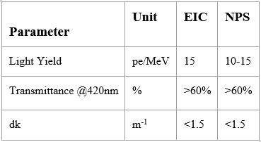
In the following sections, we describe our progress toward characterizing 28 crystals with dimensions 20x2x2 cm from Shanghai Institute of Ceramics Chinese Academy of Sciences (SICCAS), received in December 2015. Among the parameters measured were the crystals’ longitudinal transmittances, their transverse transmittances, and their light yield as a function of gate width in order to identify the fast as well as slow luminescence components. Our aim is to identify crystals that are suitable for use in the EIC and NPS, using the following parameters as guidelines:
Another relevant issue to the use of PWO in electromagnetic calorimeters is radiation hardness, which can be quantified by calculating induced absorption coefficients (dk values). As technology progresses, this factor has become even more of a concern as crystal calorimeters are being exposed to higher luminosities and higher center of mass energies than ever before (Zhu 63). Radiation damage occurs under high energy environments due to deformations that occur in the crystal lattice. Matrix host ions are displaced from their lattice site, creating vacancies that can be occupied by free electrons. Such a deformation is called a color center (Borisevitch 2013). The electrons within color centers absorb light, thus decreasing optical transmittance and making the crystal less transparent to its own scintillation light. Over time, this can degrade the energy resolution of the detector. Thus, transverse transmittances were measured prior to and after irradiation.
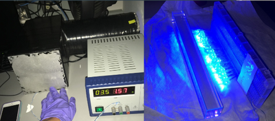
Fortunately, there are ways to restore crystal optical properties, though not all of them are practical while the crystals are in the array. It is possible to remove color centers by a process called thermal annealing, which involves heating the crystals to a temperature above 200 degrees Celsius (Borisevitch 2013). This is because the spontaneous relaxation of color centers has an exponential correlation with temperature. While this is effective, it is impractical while the calorimeter is assembled because it would be time consuming, it would take a lot of energy, and such a curing system would not be able to run in situ with data collection (Borisevitch 2013). What has instead been proposed as a solution is "optical bleaching" through LED curing, which would provide enough energy to excite electrons and release them from color centers, restoring optical transparency (Borisevitch 2013). Such a system could be more practical as it wouldn't require manipulating crystal temperature. Thus, after taking post-irradiation transverse transmittance measurements, crystals were subsequently placed into a blue LED curing box lined with millipore (a reflective material) to determine whether or not transmittance would be improved after LED treatment.
Future Projects Involving PWO
The upcoming Electron Ion Collider project will focus on providing a 3-dimensional image of nucleon to better understand sea quarks and gluons and the formation of nucleon spin in the field of Quantum Chromodynamics. Generalized Parton Distributions (GPD) and Transverse Momentum-Dependent Parton Distributions (TMD) offer clear pictures of momentum tomography and spatial tomography necessary to deconstruct nucleon structure. The study of exclusive and inclusive reactions necessary to determine GPDs and TMDs requires high luminosity beams and a world-class detector. Within the detector, the PbWO4 crystals will play a pivotal role in Particle Identification (PID) and particle reconstruction and must have good angular resolution, energy resolution, and efficiency as electromagnetic calorimeters. In particular, they will potentially be located in the Barel EM Calorimeter (BEM) and …???(Huang).
It is currently projected that the NPS will contain an array including up to 1116 scintillating PWO crystals, as well as up to 208 PbF2 crystals (Horn 2015). The NPS is designed to study hard exclusive processes in order to gain new insight into the distribution of quarks and gluons within hadrons. Such information cannot be provided by previous studies examining just inclusive and elastic scattering, as such experiments are limited to one-dimensional form factors and longitudinal parton distributions, which fail to describe certain aspects of the proton’s structure such as its orbital angular momentum (Horn 2015, “The Neutral-Particle Spectrometer”). Using QCD theorems, hard exclusive processes can be described in terms of Generalized Parton Distributions (GPDs) and Transverse Momentum-Dependent Parton Distributions (TMDs), which together enable the description of a nucleon as a 3-dimensional object ("The Neutral Particle Spectrometer"). The construction of the NPS is thus at the forefront of nuclear discovery.
Methods
Light Transmittance
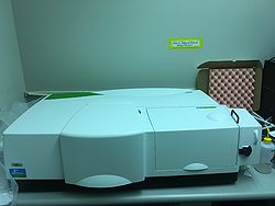
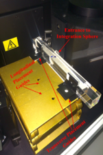
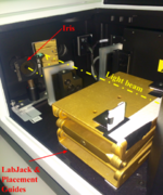
The light transmission of each crystal was measured using the Lambda 950 Spectrophotometer. The crystals were placed in the second compartment and a Labjack was installed and secured to place the crystal at the same height as the light beam. On the Labjack, fixed placement guides were put in place to ensure the constant crystal placement across all trials. An iris with a diameter of 11.5 mm was also installed and secured in the same compartment to ensure that all light from the spectrometer was entering the face of the crystal. After every five trails, the machine was auto-zeroed to ensure accuracy of measurements.
Transverse Transmission
Transverse transmission was measured with the beam firing through the middle of each 20 cm crystal. Paper was placed down to prevent scratches and the crystal was put in the placement guides. All crystals were unwrapped, cleaned with isopropanol, and marked with sharpie to indicate orientation of the crystals.
Longitudinal Transmission
To prepare the crystals for longitudinal transmission, each crystal was first wrapped in 3 layers of teflon tape and second 1 layer of electrical tape. To measure longitudinal transmittance, each crystal was placed in the corresponding placement guides.
Light Yield
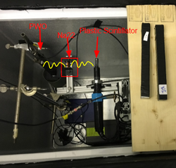
The crystals were prepared for light yield measurements by placing one layer of electrical tape over three layers of teflon tape on one open face of the crystal. The baseline gate width used was 100ns. The light yield apparatus was set up in a dark box that was temperature-controlled and set to 18 degrees Celsius. Na22, a radioactive isotope, provided two photons going in opposite directions through beta positive decay. Two scintillators, each attached to a PMT, were placed at either side of the Na22 source. A plastic trigger scintillator was placed to the right, so whenever a signal from the trigger PMT was detected, a gate was created. As a result, the only events from the crystal PMT that were read occurred at exactly the same time that the signal from the trigger PMT was detected. This means that the only events that were read were the photons coming from the Na22 source.
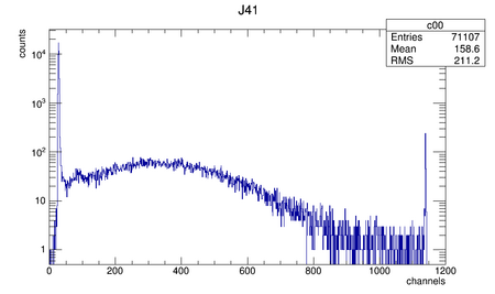
The larger, broad peak represents the crystal peak: the number of channels generated by the Photomultiplier Tube (PMT) as a result of crystal scintillation. The less broad peak to the left is the Single Electron Peak (SEP), which corresponds to the number of channels generated by the PMT as a result of a single photon hitting the crystal PMT. Each channel represents a charge of 256pC, which is the equivalent of 1.6 billion photoelectrons. The thin spike at the very left of the graph is the pedestal, which represents the "0" of the PMT. This is created due to events where a particle hit the trigger PMT but not the crystal, meaning that when the gate was open for the crystal PMT, no events of significance were read.
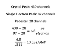
The equation to calculate light yield from this graph is provided below.
X-ray Radiation
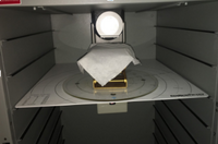
To irradiate the crystals, a Faxitron CP160 was used. It was set to 160kV and 6.3mA. A lab jack set to a height of 14.16cm was placed on shelf 8 of the machine, and the calculated ionization rate at this position was 6,970 R/min. A tissue layer was placed on top of the lab jack to protect the crystal (which was unwrapped) from scratches. When the crystal was placed into the machine, it was centered around the beam origin. A sharpie marker was used to indicate which side of the crystal was being irradiated so it was clear which side should have been facing front in the spectrometer when taking transverse transmittances before and after irradiation.
To quantify crystal radiation hardness, we used the equation for induced absorption coefficient (dk) at 420nm (the peak emission wavelength of the crystal):
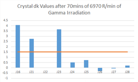
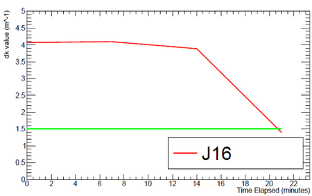
The "crystal length" value was .02m, as this was the depth of the crystal that was irradiated. Ideally, crystals would have had a dk value of 0, but to be usable in the NPS and EIC they just needed to have a dk value below 1.5.
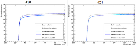
If transmittance seemed to decrease considerably post irradiation, crystals were placed inside an LED curing chamber with 34 Blue LED lights, lined with millipore. Voltage was set to 3.5V. Crystals were placed in this apparatus for periods of 7 minutes and then remeasured for transverse transmittance. If the measurement was still too low, the crystals were placed inside the LED chamber again up to three times.
Data Analysis
Longitudinal Transmittance
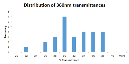
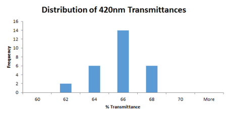
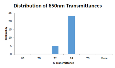
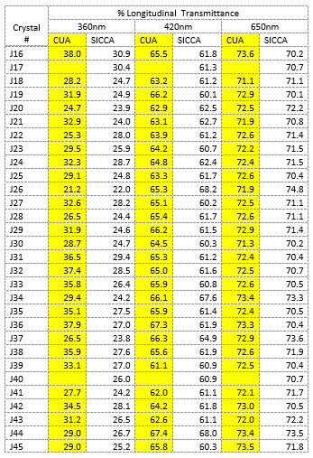
Crystals tended to vary more in longitudinal transmittance at shorter wavelengths, as can be seen from the above histograms.
Transverse Transmission/Induced Absorption Coefficients
The above graph shows that several crystal did not pass the induced absorption coefficient test (dk>1.5).
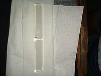
The above picture shows visible radiation damage to J23 due to the formation of color centers. The arrow indicates the side that the crystal was irradiated on.
Optical Bleaching
After 21 minutes, the dk value of J16 was lowered to below 1.5 (the threshold to be considered "usable").
Light Yield
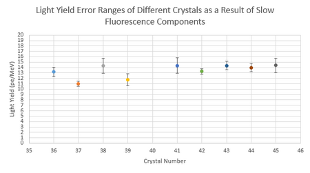
Error bars show 1 standard deviation from the mean Light Yield value. The mean represents the average light yield obtained using data at different gate widths (200ns, 300ns, 400ns, 500ns, 600ns, 700ns, 800ns, and 900ns).
Conclusion
Many of the crystal samples passed longitudinal/transverse transmittance tests prior to irradiation (>60% at 420nm), but a few failed transverse transmittance tests post irradiation and had dk values below the acceptable range. The blue LED curing system showed some promise in restoring optical properties, but not enough data was taken to draw any conclusions. Another concern that was raised is that July/August measurements for light yield seemed to be a lot lower than measurements taken earlier (around May). As consequence, many crystals did not fall within the acceptable light yield range for use in the EIC (>15 pe/MeV).
In response, more testing is needed to examine all potential variables that could be causing this variation. The high humidity due to the season could have been playing a role, even though Lead Tungstate is not particularly hydroscopic. It has been established that Lead Tungstate crystals are temperature sensitive (a temperature increase of 1 degree Celsius can decrease light yield by 2%), but the light yield apparatus is temperature controlled and measurements were not taken until the temperature of the box and crystals was 18 degrees Celsius. Seeing variance in light yield measurements could indicate inaccurate temperature readings. Gamma irradiation's effect on light yield is also something that needs to be examined. It could also be due to internal machinery being exposed to lots of heat and humidity over the past month, which may have impacted how well the setup functions.
Extensions of this project will include testing whether are not infrared LED lights would be more practical for use in a curing system, as infrared lights are less invasive and the curing system could run concurrently with data collection. This would probably not be possible with a blue LED curing system. Further, relationships between different crystal properties will continue to be investigated, such as the relationship between transverse transmittance and light yield, or the relationship between radiation hardness and light yield. This would lead to more efficient characterization as taking one measurement could give insight into expected values for other measurements.
8/5/16 Presentation
File:Characterization of Lead Tungstate Crystals for Neutral Pion Detection.pdf
Works Cited
Borisevitch, Andrei E., Valeri I. Dormenev, Andrei A. Fedorov, Mikhail V. Korjik, Till Kuske, Vitali Mechinsky, Oleg V. Missevitch, Rainer W. Novotny, Rodger Rusack, and Alexander V. Singovski. "Stimulation of Radiation Damage Recovery of Lead Tungstate Scintillation Crystals Operating in a High Dose-Rate Radiation Environment." IEEE Trans. Nucl. Sci. IEEE Transactions on Nuclear Science 60.2 (2013): 1368-372. Print.
Horn, Tanja. "A PbWO 4 -based Neutral Particle Spectrometer in Hall C at 12 GeV JLab." J. Phys.: Conf. Ser. Journal of Physics: Conference Series 587 (2015): 012048. Print.
Novotny, R., W. Doring, K. Mengel, V. Metag, and C. Pienne. "Response of a PbWO/sub 4/-scintillator Array to Electrons in the Energy Regime below 1 GeV." 1996 IEEE Nuclear Science Symposium. Conference Record 44.3 (1997): 477-83. Print.
"Neutral-Particle Spectrometer." (2014): n. pag. Web. <https://phys.cst.temple.edu/qcd/doc/NPS_WP_11262014_final.pdf>.
Huang, H. Z., and C. Woody. "EIC Detector R&D Progress Report." Electron Ion Collider RSS. N.p., 15 June 2016. Web. 08 Aug. 2016.
Zhu, Ren-Yuan. "Precision Crystal Calorimeters in High Energy Physics: Past, Present and Future." AIP Conference Proceedings (2006): n. pag. Web.
Note: All crystal measurements and specifics can also be found in the Crystal Log on the Neutral Particle Spectrometer page
